

|
| 产地 | 进口、国产 |
| 品牌 | 上海莼试 |
| 保存条件 | Store at -20 °C |
| 货号 | CS10630 |
| 应用范围 | WB=1:100-500 ELISA=1:500-1000 IP=1:20-100 IHC-P=1:100-500 IHC-F=1:100-500 ICC=1:100-500 IF=1:100-500 |
| CAS编号 | |
| 抗体名 | Anti-Acetyl-p53(K382) |
| 克隆性 | |
| 靶点 | 详见说明书 |
| 适应物种 | 详见说明书 |
| 形态 | 详见说明书 |
| 宿主 | 详见说明书 |
| 亚型 | IgG |
| 标识物 | 详见说明书 |
| 浓度 | 1mg/1ml% |
| 免疫原 | KLH conjugated Synthesised acetylpeptide derived from human p53 around the acetylation site of K382 |
产品订购信息:
英文名称 Anti-Acetyl-p53(K382)
中文名称 乙酰化P53(Lys382)抗体规格
别 名 p53 (Acetyl K382); p53 (Acetyl Lys382); Acetyl-p53 (Lys382); Widespread p53; Wtp53; Antigen NY-CO-13; Cellular tumor antigen p53; Cys 51 Stop; HGNC11998; LFS1; p53; p53 Cellular Tumor Antigen; p53 Tumor Suppressor; Phosphoprotein p53; TP53; Transformation related protein 53; TRP53; Tumor protein p53; Tumour Protein p53; P53_HUMAN; TP53.

浓 度 1mg/1ml
规 格 0.1ml/100μg 0.2ml/200μg
抗体来源 Rabbit
克隆类型 polyclonal
交叉反应 Human
产品类型 一抗 乙酰化抗体
研究领域 细胞生物 神经生物学 信号转导 细胞凋亡
蛋白分子量 predicted molecular weight: 43kDa
性 状 Lyophilized or Liquid
免 疫 原 KLH conjugated Synthesised acetylpeptide derived from human p53 around the acetylation site of K382
亚 型 IgG
纯化方法 affinity purified by Protein A
储 存 液 0.01M PBS, pH 7.4 with 10 mg/ml BSA and 0.1% Sodium azide
乙酰化P53(Lys382)抗体规格 产品应用 WB=1:100-500 ELISA=1:500-1000 IP=1:20-100 IHC-P=1:100-500 IHC-F=1:100-500 ICC=1:100-500 IF=1:100-500
(石蜡切片需做抗原修复)
not yet tested in other applications.
optimal dilutions/concentrations should be determined by the end user.
保存条件 Store at -20 °C for one year. Avoid repeated freeze/thaw cycles. The lyophilized antibody is stable at room temperature for at least one month and for greater than a year when kept at -20°C. When reconstituted in sterile pH 7.4 0.01M PBS or diluent of antibody the antibody is stable for at least two weeks at 2-4 °C.
Important Note This product as supplied is intended for research use only, not for use in human, therapeutic or diagnostic applications.
产品介绍 This gene encodes a tumor suppressor protein containing transcriptional activation, DNA binding, and oligomerization domains. The encoded protein responds to diverse cellular stresses to regulate expression of target genes, thereby inducing cell cycle arrest, apoptosis, senescence, DNA repair, or changes in metabolism. Mutations in this gene are associated with a variety of human cancers, including hereditary cancers such as Li-Fraumeni syndrome. Alternative splicing of this gene and the use of alternate promoters result in multiple transcript variants and isoforms. Additional isoforms have also been shown to result from the use of alternate translation initiation codons (PMIDs: 12032546, 20937277). [provided by RefSeq, Feb 2013].
Function : Acts as a tumor suppressor in many tumor types; induces growth arrest or apoptosis depending on the physiological circumstances and cell type. Involved in cell cycle regulation as a trans-activator that acts to negatively regulate cell division by controlling a set of genes required for this process. One of the activated genes is an inhibitor of cyclin-dependent kinases. Apoptosis induction seems to be mediated either by stimulation of BAX and FAS antigen expression, or by repression of Bcl-2 expression. Implicated in Notch signaling cross-over. Prevents CDK7 kinase activity when associated to CAK complex in response to DNA damage, thus stopping cell cycle progression. Isoform 2 enhances the transactivation activity of isoform 1 from some but not all TP53-inducible promoters. Isoform 4 suppresses transactivation activity and impairs growth suppression mediated by isoform 1. Isoform 7 inhibits isoform 1-mediated apoptosis.
Subunit : Interacts with AXIN1. Probably part of a complex consisting of TP53, HIPK2 and AXIN1 (By similarity). Binds DNA as a homotetramer. Interacts with histone acetyltransferases EP300 and methyltransferases HRMT1L2 and CARM1, and recruits them to promoters. In vitro, the interaction of TP53 with cancer-associated/HPV (E6) viral proteins leads to ubiquitination and degradation of TP53 giving a possible model for cell growth regulation. This complex formation requires an additional factor, E6-AP, which stably associates with TP53 in the presence of E6. Interacts (via C-terminus) with TAF1; when TAF1 is part of the TFIID complex. Interacts with ING4; this interaction may be indirect. Found in a complex with CABLES1 and TP73. Interacts with HIPK1, HIPK2, and TP53INP1. Interacts with WWOX. May interact with HCV core protein. Interacts with USP7 and SYVN1. Interacts with HSP90AB1. Interacts with CHD8; leading to recruit histone H1 and prevent transactivation activity (By similarity). Interacts with ARMC10, BANP, CDKN2AIP, NUAK1, STK11/LKB1, UHRF2 and E4F1. Interacts with YWHAZ; the interaction enhances TP53 transcriptional activity. Phosphorylation of YWHAZ on 'Ser-58' inhibits this interaction. Interacts (via DNA-binding domain) with MAML1 (via N-terminus). Interacts with MKRN1. Interacts with PML (via C-terminus). Interacts with MDM2; leading to ubiquitination and proteasomal degradation of TP53. Directly interacts with FBXO42; leading to ubiquitination and degradation of TP53. Interacts (phosphorylated at Ser-15 by ATM) with the phosphatase PP2A-PPP2R5C holoenzyme; regulates stress-induced TP53-dependent inhibition of cell proliferation. Interacts with PPP2R2A. Interacts with AURKA, DAXX, BRD7 and TRIM24. Interacts (when monomethylated at Lys-382) with L3MBTL1. Isoform 1 interacts with isoform 2 and with isoform 4. Interacts with GRK5. Binds to the CAK complex (CDK7, cyclin H and MAT1) in response to DNA damage. Interacts with CDK5 in neurons. Interacts with AURKB, UHRF2 and NOC2L. Interacts (via N-terminus) with PTK2/FAK1; this promotes ubiquitination by MDM2. Interacts with PTK2B/PYK2; this promotes ubiquitination by MDM2. Interacts with PRKCG. Interacts with human cytomegalovirus/HHV-5 protein UL123.
Subcellular Location : Cytoplasm. Nucleus. Nucleus, PML body. Endoplasmic reticulum. Note=Interaction with BANP promotes nuclear localization. Recruited into PML bodies together with CHEK2.
Isoform 2: Nucleus. Cytoplasm. Note=Localized mainly in the nucleus with minor staining in the cytoplasm.
Isoform 3: Nucleus. Cytoplasm. Note=Localized in the nucleus in most cells but found in the cytoplasm in some cells.
Isoform 4: Nucleus. Cytoplasm. Note=Predominantly nuclear but translocates to the cytoplasm following cell stress.
Isoform 7: Nucleus. Cytoplasm. Note=Localized mainly in the nucleus with minor staining in the cytoplasm.
Isoform 8: Nucleus. Cytoplasm. Note=Localized in both nucleus and cytoplasm in most cells. In some cells, forms foci in the nucleus that are different from nucleoli.
Isoform 9: Cytoplasm.
Tissue Specificity : Ubiquitous. Isoforms are expressed in a wide range of normal tissues but in a tissue-dependent manner. Isoform 2 is expressed in most normal tissues but is not detected in brain, lung, prostate, muscle, fetal brain, spinal cord and fetal liver. Isoform 3 is expressed in most normal tissues but is not detected in lung, spleen, testis, fetal brain, spinal cord and fetal liver. Isoform 7 is expressed in most normal tissues but is not detected in prostate, uterus, skeletal muscle and breast. Isoform 8 is detected only in colon, bone marrow, testis, fetal brain and intestine. Isoform 9 is expressed in most normal tissues but is not detected in brain, heart, lung, fetal liver, salivary gland, breast or intestine.
Post-translational modifications : Acetylated. Acetylation of Lys-382 by CREBBP enhances transcriptional activity. Deacetylation of Lys-382 by SIRT1 impairs its ability to induce proapoptotic program and modulate cell senescence.
Phosphorylation on Ser residues mediates transcriptional activation. Phosphorylated by HIPK1. Phosphorylation at Ser-9 by HIPK4 increases repression activity on BIRC5 promoter. Phosphorylated on Thr-18 by VRK1. Phosphorylated on Ser-20 by CHEK2 in response to DNA damage, which prevents ubiquitination by MDM2. Phosphorylated on Ser-20 by PLK3 in response to reactive oxygen species (ROS), promoting p53/TP53-mediated apoptosis. Phosphorylated on Thr-55 by TAF1, which promotes MDM2-mediated degradation. Phosphorylated on Ser-33 by CDK7 in a CAK complex in response to DNA damage. Phosphorylated on Ser-46 by HIPK2 upon UV irradiation. Phosphorylation on Ser-46 is required for acetylation by CREBBP. Phosphorylated on Ser-392 following UV but not gamma irradiation. Phosphorylated upon DNA damage, probably by ATM or ATR. Phosphorylated on Ser-15 upon ultraviolet irradiation; which is enhanced by interaction with BANP. Phosphorylated by NUAK1 at Ser-15 and Ser-392; was initially thought to be mediated by STK11/LKB1 but it was later shown that it is indirect and that STK11/LKB1-dependent phosphorylation is probably mediated by downstream NUAK1 (PubMed:21317932). It is unclear whether AMP directly mediates phosphorylation at Ser-15. Phosphorylated on Thr-18 by isoform 1 and isoform 2 of VRK2. Phosphorylation on Thr-18 by isoform 2 of VRK2 results in a reduction in ubiquitination by MDM2 and an increase in acetylation by EP300. Stabilized by CDK5-mediated phosphorylation in response to genotoxic and oxidative stresses at Ser-15, Ser-33 and Ser-46, leading to accumulation of p53/TP53, particularly in the nucleus, thus inducing the transactivation of p53/TP53 target genes. Phosphorylated at Ser-315 and Ser-392 by CDK2 in response to DNA-damage.
Dephosphorylated by PP2A-PPP2R5C holoenzyme at Thr-55. SV40 small T antigen inhibits the dephosphorylation by the AC form of PP2A.
May be O-glycosylated in the C-terminal basic region. Studied in EB-1 cell line.
Ubiquitinated by MDM2 and SYVN1, which leads to proteasomal degradation. Ubiquitinated by RFWD3, which works in cooperation with MDM2 and may catalyze the formation of short polyubiquitin chains on p53/TP53 that are not targeted to the proteasome. Ubiquitinated by MKRN1 at Lys-291 and Lys-292, which leads to proteasomal degradation. Deubiquitinated by USP10, leading to its stabilization. Ubiquitinated by TRIM24, which leads to proteasomal degradation. Ubiquitination by TOPORS induces degradation. Deubiquitination by USP7, leading to stabilization. Isoform 4 is monoubiquitinated in an MDM2-independent manner.
Monomethylated at Lys-372 by SETD7, leading to stabilization and increased transcriptional activation. Monomethylated at Lys-370 by SMYD2, leading to decreased DNA-binding activity and subsequent transcriptional regulation activity. Lys-372 monomethylation prevents interaction with SMYD2 and subsequent monomethylation at Lys-370. Dimethylated at Lys-373 by EHMT1 and EHMT2. Monomethylated at Lys-382 by SETD8, promoting interaction with L3MBTL1 and leading to repress transcriptional activity. Demethylation of dimethylated Lys-370 by KDM1A prevents interaction with TP53BP1 and represses TP53-mediated transcriptional activation.
Sumoylated by SUMO1.
DISEASE : Note=TP53 is found in increased amounts in a wide variety of transformed cells. TP53 is frequently mutated or inactivated in about 60% of cancers. TP53 defects are found in Barrett metaplasia a condition in which the normally stratified squamous epithelium of the lower esophagus is replaced by a metaplastic columnar epithelium. The condition develops as a complication in approximately 10% of patients with chronic gastroesophageal reflux disease and predisposes to the development of esophageal adenocarcinoma.
Defects in TP53 are a cause of esophageal cancer (ESCR) [MIM:133239].
Defects in TP53 are a cause of Li-Fraumeni syndrome (LFS) [MIM:151623]. LFS is an autosomal dominant familial cancer syndrome that in its classic form is defined by the existence of a proband affected by a sarcoma before 45 years with a first degree relative affected by any tumor before 45 years and another first degree relative with any tumor before 45 years or a sarcoma at any age. Other clinical definitions for LFS have been proposed (PubMed:8118819 and PubMed:8718514) and called Li-Fraumeni like syndrome (LFL). In these families affected relatives develop a diverse set of malignancies at unusually early ages. Four types of cancers account for 80% of tumors occurring in TP53 germline mutation carriers: breast cancers, soft tissue and bone sarcomas, brain tumors (astrocytomas) and adrenocortical carcinomas. Less frequent tumors include choroid plexus carcinoma or papilloma before the age of 15, rhabdomyosarcoma before the age of 5, leukemia, Wilms tumor, malignant phyllodes tumor, colorectal and gastric cancers.
Defects in TP53 are involved in head and neck squamous cell carcinomas (HNSCC)
Defects in TP53 are a cause of lung cancer (LNCR) [MIM:211980]. LNCR is a common malignancy affecting tissues of the lung. The most common form of lung cancer is non-small cell lung cancer (NSCLC) that can be divided into 3 major histologic subtypes: squamous cell carcinoma, adenocarcinoma, and large cell lung cancer. NSCLC is often diagnosed at an advanced stage and has a poor prognosis.
Defects in TP53 are a cause of choroid plexus papilloma (CPLPA) [MIM:260500]. Choroid plexus papilloma is a slow-growing benign tumor of the choroid plexus that often invades the leptomeninges. In children it is usually in a lateral ventricle but in adults it is more often in the fourth ventricle. Hydrocephalus is common, either from obstruction or from tumor secretion of cerebrospinal fluid. If it undergoes malignant transformation it is called a choroid plexus carcinoma. Primary choroid plexus tumors are rare and usually occur in early childhood.
Defects in TP53 are a cause of adrenocortical carcinoma (ADCC) [MIM:202300]. ADCC is a rare childhood tumor of the adrenal cortex. It occurs with increased frequency in patients with the Beckwith-Wiedemann syndrome and is a component tumor in Li-Fraumeni yndrome.
Similarity : Belongs to the p53 family.
Database links : NCBI Reference Sequence: NP_001119584.1
UniProtKB/Swiss-Prot: P04637.4
wtp53广泛的研究发现P53。P53蛋白水平在正常细胞中表达低,在DNA突变时或各种各样细胞遇难信号时反应增加。该基因突变或缺失是导致许多发生的原因。
野生型P53(wt-p53)可诱导细胞凋亡,并通过细胞凋亡抑制,而P53的突变或缺失则可抑制野生型P53的功能,使得缺陷细胞得以存活下来,从而导致发生。 P53同时也是细胞凋亡的调控因子。此抗体可用于P53的研究。

Anti-DMT1/FITC 荧光素标记金属离子转运体1抗体IgGMulti-class antibodies规格: 0.2ml
Anti-Phospho-Mcl1 (Ser159/Thr163)/FITC 荧光素标记磷酸化髓样细胞-1抗体IgGMulti-class antibodies规格: 0.2ml
Rhesus antibody Rh CACH2/CaV1.2 L型钙通道蛋白抗体 规格 0.1ml
C-Mer/MERTK (proto-oncogene tyrosine kinase) c-mer原癌基因酪酸激酶抗原 0.5mg
HSPA6 英文名称: 热休克蛋白70家族蛋白6抗体 0.2ml
Rhesus antibody Rh Sema6A Sema6A抗体 规格 0.1ml
Anti-Phospho-Mcl1 (Ser159/Thr163)/FITC 荧光素标记磷酸化髓样细胞-1抗体IgGMulti-class antibodies规格: 0.2ml
CAM(Human calmodulin) ELISA Kit 人钙调素Multi-class antibodies规格: 48T
Anti-Phospho-Stathmin (Ser38) 磷酸化原癌基因蛋白18Multi-class antibodies规格: 0.1ml
Rhesus antibody Rh Lactoferrin 乳铁蛋白抗体 规格 0.1ml
GM-CSF ELISA Kit 大鼠粒细胞巨噬细胞集落刺激因子 96T
PPP2R1A 英文名称: 蛋白质磷酸酶2调节亚基1A抗体 0.2ml
Contactin 4 + 6 英文名称: 轴突相关粘附分子抗体 0.2ml
Anti-Phospho-Stathmin (Ser38) 磷酸化原癌基因蛋白18Multi-class antibodies规格: 0.1ml
MYS(Human Myosin) ELISA Kit 人肌球蛋白Multi-class antibodies规格: 48T
Anti-PAF 血小板活化因子抗体Multi-class antibodies规格: 0.1ml
Rhesus antibody Rh HPV18 E6 monoclonal 人类状瘤病毒18 E6单克隆抗体 规格 0.2ml
GnRH ELISA Kit 大鼠激素释放激素 96T
Phospho-PBK (Thr9) 英文名称: 磷酸化PDZ连接激酶/T-LAK细胞源蛋白激酶抗体 0.1ml
CDC45L 英文名称: 细胞分裂周期调控蛋白45抗体 0.2ml
Anti-PAF 血小板活化因子抗体Multi-class antibodies规格: 0.1ml
CL-0264C918(人眼脉络细胞)5×10⁶cells/瓶×2
HA Others H4N8 甲型 H4N8 (A/chicken/Alabama/1/1975) 血凝素 (Hemagglutinin / HA) 人细胞裂解液 (阳性对照)
小鼠上皮细胞完全培养基 100mL
NCTC clone 929 [L cell, L-929, derivative of Sain L]小鼠成纤维细胞 NCTC clone 929 [L cell, L-929, derivative of Sain L] mouse fibroblast cells MEM(NaHCO3 1.5g/L, Sodium Pyruvate 0.11g/L)+10%FBS
IL1B Protein Human 重组人 IL-1 beta / IL1B 蛋白
PC-12(大鼠肾上腺嗜铬细胞瘤细胞) 5×10⁶cells/瓶×2 RSC96(大鼠雪旺细胞)
CL-0261BC-022(人癌细胞)5×10⁶cells/瓶×2
IFNB1 Others Mouse 小鼠 IFNB1 / IFN-beta / Ierferon beta 人细胞裂解液 (阳性对照)
人髓核细胞cDNAHNPC cDNA
T47D细胞,人管癌细胞 猪肾细胞系,IBRS-2细胞 Many types of cells包装:5 × 10⁵次方(1ml)
MC3T3-E1(小鼠胚胎成骨细胞前体细胞) 5×10⁶cells/瓶×2
HCMEC Pellet 人微内皮细胞团块 > 1 mio.cells 纤维母细胞粘附检测试剂盒Fibro
乙酰化P53(Lys382)抗体规格 JAM2 Others Rat 大鼠 JAM-2 / JAM-B 人细胞裂解液 (阳性对照)
人成纤维细胞总RNAHPrF NA
人颅盖造骨细胞完全培养基 100mL
CHO-K1细胞,中国仓鼠仓鼠卵巢细胞亚株 MC3T3-E1 Subclone 14(小鼠原成骨细胞) 小鼠细胞;CT26.WT [CT26WT]
CSF2 Protein Human 重组人 GM-CSF / CSF2 蛋白 (Fc 标签)
U-2 OS(人细胞) 5×10⁶cells/瓶×2 小鼠胚胎成纤维细胞;3T6-Swiss albino

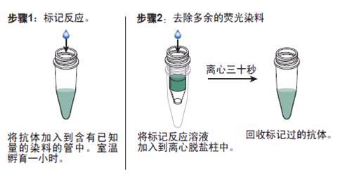
抗体的生物素化标记实验要点:
1. 乙酰化P53(Lys382)抗体规格 如在反应混合液中有叠氮钠或游离氨基存在,会抑制标记反应。因此,蛋白质在反应前要对 0.1mol/L碳酸氢钠缓冲液或0.5mol/L硼酸缓冲液充分透析;
2.所用的NHSB及待生物素化蛋白质之间的分子比按蛋白质表面的ε-氨基的密度会有所不同,选择不当则影响标记的效率,应先用几个不同的分子比来筛选最适条件;
3.用NHSB量过量也是不利的,抗原的结合位点可能因此被封闭,导致抗体失活;
4.由于抗体的氨基不易接近可能造成生物素化不足,此时可加入去污剂如 Triton x-100, Tween20等;
5.当游离ε-氨基(赖氨酸残基的氨基)存在于抗体的抗原结合位点时,或位于酶的催化位点时,生物素化会降低或损伤抗体蛋白的结合力或活性;
6.生物素还可能与不同的功能基团,如羰基、氨基、巯基、异咪唑基及*基,也可与糖基共价结合;
7.交联反应后,应充分透析,否则,残余的生物素会对生物素化抗体与亲和素的结合产生竞争作用;
8.在细胞的荧光标记实验中,中和亲和素的本底低,但由于链霉亲和素含有少量正电荷,故对某些细胞可导致高本底。
抗体的鉴定:
1)乙酰化P53(Lys382)抗体规格 抗体的效价鉴定:不管是用于诊断还是用于,制备抗体的目的都是要求较高效价。不同的抗原制备的抗体,要求的效价不一。鉴定效价的方法很多,包括有试管凝集反应,琼脂扩散试验,酶联免疫吸附试验等。常用的抗原所制备的抗体一般都有约成的鉴定效价的方法,以资比较。如制备抗抗体的效价,一般就采用琼脂扩散试验来鉴定。
2)抗体的特异性鉴定:抗体的特异性是指与相应抗原或近似抗原物质的识别能力。抗体的特异性高,它的识别能力就强。衡量特异性通常以交叉反应率来表示。交叉反应率可用竞争抑制试验测定。以不同浓度抗原和近似抗原分别做竞争抑制曲线,计算各自的结合率,求出各自在IC50时的浓度,并按公式计算交叉反应率。
如果所用抗原浓度IC50浓度为pg/管,而一些近似抗原物质的IC50浓度几乎是无穷大时,表示这一抗血清与其他抗原物质的交叉反应率近似为0,即该血清的特异性较好。
3)抗体亲和力:是指抗体和抗原结合的牢固程度。亲和力的高低是由抗原分子的大小,抗体分子的结合位点与抗原决定簇之间立体构型的合适度决定的。有助于维持抗原抗体复合物稳定的分子间力有氢键,疏水键,侧链相反电荷基因的库仑力,范德华力和空间斥力。亲和力常以亲和常数K表示,K的单位是L/mol。抗体亲和力的测定对抗体的筛选,确定抗体的用途,验证抗体的均一性等均有重要意义。