

|
| 产地 | 进口、国产 |
| 品牌 | 上海莼试 |
| 保存条件 | Store at -20 °C |
| 货号 | CS10685 |
| 应用范围 | WB=1:100-500 ELISA=1:500-1000 IP=1:20-100 IHC-P=1:100-500 IHC-F=1:100-500 IF=1:100-500 |
| CAS编号 | |
| 抗体名 | Anti-Polycystin 1 |
| 克隆性 | |
| 靶点 | 详见说明书 |
| 适应物种 | 详见说明书 |
| 形态 | 详见说明书 |
| 宿主 | 详见说明书 |
| 亚型 | IgG |
| 标识物 | 详见说明书 |
| 浓度 | 1mg/1ml% |
| 免疫原 | KLH conjugated synthetic peptide derived from human Polycystin 1 N-terminus |
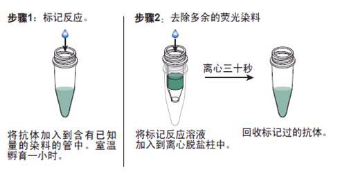
抗体的生物素化标记实验要点:
1. 蛋白1抗体费用 如在反应混合液中有叠氮钠或游离氨基存在,会抑制标记反应。因此,蛋白质在反应前要对 0.1mol/L碳酸氢钠缓冲液或0.5mol/L硼酸缓冲液充分透析;
2.所用的NHSB及待生物素化蛋白质之间的分子比按蛋白质表面的ε-氨基的密度会有所不同,选择不当则影响标记的效率,应先用几个不同的分子比来筛选最适条件;
3.用NHSB量过量也是不利的,抗原的结合位点可能因此被封闭,导致抗体失活;
4.由于抗体的氨基不易接近可能造成生物素化不足,此时可加入去污剂如 Triton x-100, Tween20等;
5.当游离ε-氨基(赖氨酸残基的氨基)存在于抗体的抗原结合位点时,或位于酶的催化位点时,生物素化会降低或损伤抗体蛋白的结合力或活性;
6.生物素还可能与不同的功能基团,如羰基、氨基、巯基、异咪唑基及*基,也可与糖基共价结合;
7.交联反应后,应充分透析,否则,残余的生物素会对生物素化抗体与亲和素的结合产生竞争作用;
8.在细胞的荧光标记实验中,中和亲和素的本底低,但由于链霉亲和素含有少量正电荷,故对某些细胞可导致高本底。
产品订购信息:
英文名称 Anti-Polycystin 1
中文名称 蛋白1抗体费用
别 名 Autosomal dominant polycystic kidney disease protein 1; PBP; PKD; PKD1; Polycystic Kidney Disease 1; Polycystin 1 Precursor; PKD1; Pc-1; TRPP1; PKD1_HUMAN.

浓 度 1mg/1ml
规 格 0.2ml/200μg
抗体来源 Rabbit
克隆类型 polyclonal
交叉反应 Human, Mouse, Dog
产品类型 一抗
研究领域 细胞生物 免疫学 发育生物学
蛋白分子量 predicted molecular weight: 460kDa
性 状 Lyophilized or Liquid
免 疫 原 KLH conjugated synthetic peptide derived from human Polycystin 1 N-terminus
亚 型 IgG
纯化方法 affinity purified by Protein A
储 存 液 0.01M PBS, pH 7.4 with 10 mg/ml BSA and 0.1% Sodium azide
蛋白1抗体费用 产品应用 WB=1:100-500 ELISA=1:500-1000 IP=1:20-100 IHC-P=1:100-500 IHC-F=1:100-500 IF=1:100-500
(石蜡切片需做抗原修复)
not yet tested in other applications.
optimal dilutions/concentrations should be determined by the end user.
保存条件 Store at -20 °C for one year. Avoid repeated freeze/thaw cycles. The lyophilized antibody is stable at room temperature for at least one month and for greater than a year when kept at -20°C. When reconstituted in sterile pH 7.4 0.01M PBS or diluent of antibody the antibody is stable for at least two weeks at 2-4 °C.
Important Note This product as supplied is intended for research use only, not for use in human, therapeutic or diagnostic applications.
产品介绍 This gene encodes a member of the polycystin protein family. The encoded glycoprotein contains a large N-terminal extracellular region, multiple transmembrane domains and a cytoplasmic C-tail. It is an integral membrane protein that functions as a regulator of calcium permeable cation channels and intracellular calcium homoeostasis. It is also involved in cell-cell/matrix interactions and may modulate G-protein-coupled signal-transduction pathways. It plays a role in renal tubular development, and mutations in this gene cause autosomal dominant polycystic kidney disease type 1 (ADPKD1). ADPKD1 is characterized by the growth of fluid-filled cysts that replace normal renal tissue and result in end-stage renal failure. Splice variants encoding different isoforms have been noted for this gene. Also, six pseudogenes, closely linked in a known duplicated region on chromosome 16p, have been described. [provided by RefSeq].
Function : Involved in renal tubulogenesis. Involved in fluid-flow mechanosensation by the primary cilium in renal epithelium (By similarity). Acts as a regulator of cilium length, together with PKD2 (By similarity). The dynamic control of cilium length is essential in the regulation of mechanotransductive signaling. The cilium length response creates a negative feedback loop whereby fluid shear-mediated deflection of the primary cilium, which decreases intracellular cAMP, leads to cilium shortening and thus decreases flow-induced signaling (By similarity). May be an ion-channel regulator. Involved in adhesive protein-protein and protein-carbohydrate interactions.
Subunit : Interacts with PKD2. Interacts with PRKX; involved in differentiation and controlled morphogenesis of the kidney. Interacts with NPHP1 (via SH3 domain).
Subcellular Location : Membrane; Multi-pass membrane protein. Cell projection, cilium (By similarity). Note=PKD1 localization to the plasma and ciliary membranes requires PKD2, is independent of PKD2 channel activity, and involves stimulation of PKD1 autoproteolytic cleavage at the GPS domain.
DISEASE : Defects in PKD1 are the cause of polycystic kidney disease autosomal dominant type 1 (ADPKD1) [MIM:173900]. ADPKD is characterized by progressive formation and enlargement of cysts in both kidneys, typically leading to end-stage renal disease in adult life. Cysts also occurs in the liver and other organs. Its prevalence is estimated at about 1/1000. [SIMILARITY] Belongs to the polycystin family.
Similarity : Contains 1 C-type lectin domain.
Contains 1 GPS domain.
Contains 1 LDL-receptor class A domain.
Contains 2 LRR (leucine-rich) repeats.
Contains 1 LRRCT domain.
Contains 1 LRRNT domain.
Contains 17 PKD domains.
Contains 1 PLAT domain.
Contains 1 REJ domain.
Contains 1 WSC domain.
Database links : UniProtKB/Swiss-Prot: P98161.3
(polycystic kidney disease)为遗传性疾病,是*一种先天性异常。双侧*皮髓质均可累及,但在程度上可不同。在遗传方式上表现为常染色体显性和常染色体隐性遗传两种。
囊内上皮细胞异常增殖是ADPKD的显著特特之一,处于一种成熟不完全或重发育状态,高度提示为细胞的发育成熟调控出现障碍,使细胞处于一种未成熟状态,从而显示强增殖性。表现为细胞转运密切相关的Na+-K+-ATP ase的亚单位组合,分布及活性表达的改变;细胞信号传导异常以及离子转运通道的变化。细胞外基质异常增生是ADPKD第三种显著特征。目前许多研究已证明:这些异常均有与细胞生长有关的活性因子的参与。但关键的异常环节和途径尚未明了。因基因缺陷而致的细胞生长改变和间质形成异常,是本病的重要机制之一。

抗体的鉴定:
1)蛋白1抗体费用 抗体的效价鉴定:不管是用于诊断还是用于,制备抗体的目的都是要求较高效价。不同的抗原制备的抗体,要求的效价不一。鉴定效价的方法很多,包括有试管凝集反应,琼脂扩散试验,酶联免疫吸附试验等。常用的抗原所制备的抗体一般都有约成的鉴定效价的方法,以资比较。如制备抗抗体的效价,一般就采用琼脂扩散试验来鉴定。
2)抗体的特异性鉴定:抗体的特异性是指与相应抗原或近似抗原物质的识别能力。抗体的特异性高,它的识别能力就强。衡量特异性通常以交叉反应率来表示。交叉反应率可用竞争抑制试验测定。以不同浓度抗原和近似抗原分别做竞争抑制曲线,计算各自的结合率,求出各自在IC50时的浓度,并按公式计算交叉反应率。
如果所用抗原浓度IC50浓度为pg/管,而一些近似抗原物质的IC50浓度几乎是无穷大时,表示这一抗血清与其他抗原物质的交叉反应率近似为0,即该血清的特异性较好。
3)抗体亲和力:是指抗体和抗原结合的牢固程度。亲和力的高低是由抗原分子的大小,抗体分子的结合位点与抗原决定簇之间立体构型的合适度决定的。有助于维持抗原抗体复合物稳定的分子间力有氢键,疏水键,侧链相反电荷基因的库仑力,范德华力和空间斥力。亲和力常以亲和常数K表示,K的单位是L/mol。抗体亲和力的测定对抗体的筛选,确定抗体的用途,验证抗体的均一性等均有重要意义。

Anti-Phospho-c-Fos (Se*) /FITC 荧光素标记兔抗人、大、小鼠磷酸化c-fos抗体IgGMulti-class antibodies规格: 0.2ml
Anti-IL-22BP/IL22RA2/FITC 荧光素标记白介素22受体结合蛋白抗体IgGMulti-class antibodies规格: 0.2ml
Rhesus antibody Rh CD137/TNFRSF9 坏死因子受体超家族成员9抗体 规格 0.1ml
phospho-Klf5/CKLF/Kruppel like factor 5, Human, mo, rat, horse, bovine, rabbit, pig, dog 磷酸化肠道内富含的Kruppel样因子5抗原 0.5mg
IDH3A 英文名称: 草酰琥珀酸脱羧酶α抗体 0.2ml
Rhesus antibody Rh SRFBP1 血清应答因子结合蛋白1抗体 规格 0.2ml
Anti-IL-22BP/IL22RA2/FITC 荧光素标记白介素22受体结合蛋白抗体IgGMulti-class antibodies规格: 0.2ml
human phospho-inhibitory subunit of NF-κB Alpha,pIKB Alpha ELISA kit 人磷酸化核因子κB抑制蛋白α(IKKα human ELISA kit)Multi-class antibodies规格: 96T
Anti-Phospho-SHIP2 (Tyr1135) 磷酸化蛋白酪氨酸磷酸酶2抗体Multi-class antibodies规格: 0.1ml
Rhesus antibody Rh Goat Anti-rat IgG/PE-Cy7 PE-Cy7标记的羊抗大鼠IgG 规格 0.1ml
MC Ab(Human melanocyte antibody) ELISA Kit 人黑色素细胞抗体 96T
phospho-NFATC4 (Ser168 + Ser170) 英文名称: 磷酸化T细胞激活核转录因子4抗体 0.1ml
BEND3 英文名称: BEND3蛋白抗体 0.2ml
Anti-Phospho-SHIP2 (Tyr1135) 磷酸化蛋白酪氨酸磷酸酶2抗体Multi-class antibodies规格: 0.1ml
PGAM2(Human phosphoglycerate mutase 2) ELISA Kit 人磷酸甘油酸变位酶2Multi-class antibodies规格: 48T
Anti-MYOZ/MYOZ1 MYOZ抗体Multi-class antibodies规格: 0.2ml
Rhesus antibody Rh ILVBL 乙酰乳酸合成酶蛋白抗体 规格 0.2ml
Human histone H2A-H2B-DNA autoantibody ELISA Kit 人抗组蛋白(H2A-H2B)-DNA抗体/抗二聚体-DNA抗体 96T
Phospho-P38 MAPK (Thr180 + Tyr182) 英文名称: 磷酸化-原活化蛋白激酶p38抗体(P-p38 MAPK) 0.1ml
phospho-CEBPE (Thr74) 英文名称: 磷酸化转录调节因子C/EBPε抗体 0.1ml
Anti-MYOZ/MYOZ1 MYOZ抗体Multi-class antibodies规格: 0.2ml
CL-0154MDCK(犬肾细胞)5×10⁶cells/瓶×2
IFNB1 Others Human 人 Ierferon beta / IFN-beta / IFNB CHO细胞裂解液 (阳性对照)
人肝窦内皮细胞裂解物HHSECL
小香猪皮肤细胞;SSC-S2 组织细胞细胞,U-937细胞 NUTU-19细胞,大鼠细胞株
SV40转染人成骨细胞;hFOB 1.19
HA Others H1N3 甲型 H1N3 (A/duck/NZL/160/1976) 血凝素HA1 (Hemagglutinin) 人细胞裂解液 (阳性对照)
CL-0152MDA-MB-453(人癌细胞)5×10⁶cells/瓶×2
F10 Others Human 人 Coagulation Factor X / FX / F10 杆状病毒-昆虫细胞裂解液 (阳性对照)
人滑膜细胞裂解物HSL
NCI-H838[H838]细胞,人细胞 人,A2细胞 脑内皮细胞Many types of cells包装:5 × 10⁵次方(1ml)
兔角膜后基质层成纤维细胞;RCBBF
HAVCR2 Others Human 人 TIMD3 / TIM3 / HAVCR2 人细胞裂解液 (阳性对照)
蛋白1抗体费用 IL12A&IL27B Others Human 人 IL12A & IL27B Heterodimer 人细胞裂解液 (阳性对照)
人成细胞;MG-63
人脐带血单个核细胞
CTLL-2细胞,小鼠T细胞 人细胞,9L-B细胞 人绒毛间叶成纤维细胞RNAHVMF miRNA5 μg
恒河猴皮肤细胞;RM-S1
EPHB2 Others Human 人 EphB2 人细胞裂解液 (阳性对照)